
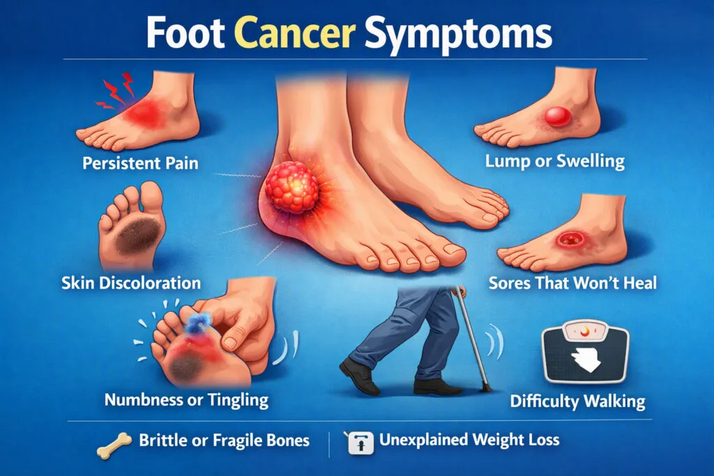
If you’ve noticed a mole that’s changed shape, a sore on your foot that won’t heal, or a dark streak under a toenail, you’re right to pay attention. Foot cancer symptoms include new or changing moles, non-healing ulcers, unexplained lumps or swelling, persistent pain, and discoloration under the nail bed. These signs are easy to dismiss as a callus, a fungal infection, or “just a bruise” — which is exactly why foot cancer is so often caught late. This guide walks through the earliest signs, what actually causes it, who’s at higher risk, and what treatment really looks like if a diagnosis is confirmed.
What Is Foot Cancer, Exactly?
“Foot cancer” isn’t one disease — it’s an umbrella term for several different cancers that can develop in the skin, soft tissue, bone, or nail unit of the foot and ankle. Some start locally; others are cancers elsewhere in the body that show up on the foot as a secondary sign. Because the feet take daily wear and tear, an abnormal spot or wound is often waved off as a routine podiatric annoyance, and that delay is where the real danger lies.
The Main Types of Foot Cancer
- Melanoma – the most serious skin cancer, and the one most associated with feet. Around half of all foot melanomas appear as a type called acral lentiginous melanoma (ALM), which often shows up on the sole or under a toenail rather than in a sun-exposed spot. Melanoma is a striking example of how a rare cancer can carry outsized risk: it accounts for only a small share of all skin cancer diagnoses overall, yet is responsible for the large majority of skin-cancer-related deaths, which is exactly why speed of diagnosis matters so much with this particular type.
- Squamous cell carcinoma (SCC) – the most common skin cancer found on the foot. It often starts as a scaly, inflamed bump that can be mistaken for a plantar wart, a callus, or a fungal patch. SCC has a well-documented link to areas of chronic irritation — old scars, long-standing ulcers, and areas of persistent inflammation are all considered higher-risk sites.
- Basal cell carcinoma (BCC) – less common on the feet, slower-growing, but still needs early treatment. It can look like a pearly bump or an open sore that doesn’t heal.
- Soft tissue sarcoma – develops in the muscle, fat, or connective tissue of the foot or ankle. It frequently starts as a painless lump, which makes it easy to ignore until it grows.
- Bone cancer (osteosarcoma and related tumors) – rarer, and more often seen in younger patients. It typically presents with swelling, deep pain, or even a fracture that happens with minimal trauma. A small number of bone cancers are linked to inherited genetic syndromes or prior radiation exposure, though for most cases no clear cause is ever identified.
- Kaposi sarcoma – linked to immune system suppression, this can appear on the feet as purple-red patches or nodules and is more likely in people who are immunocompromised.
Early Signs of Foot Cancer
The earliest indicators are usually small, painless, and easy to explain away — which is exactly why they’re worth learning to spot deliberately rather than waiting to “notice” them by accident.
The Earliest, Most Subtle Clues
- A new dark spot, or an old mole that looks slightly “off” – not necessarily dramatic, sometimes just a mole that seems to have grown a little, or gained an extra shade of color, since the last time you looked.
- A faint streak under a toenail – often mistaken for a bruise from an ill-fitting shoe, especially if you can’t recall a specific injury that would explain it.
- A small scaly or rough patch that resembles dry skin or the start of a fungal infection, but doesn’t improve with moisturizer or antifungal cream after a couple of weeks.
- A pinpoint sore that scabs, heals a little, then reopens in the same spot — a cycle that’s easy to mistake for a healing wound that keeps getting irritated by a shoe.
- A tiny, firm lump you can feel but barely see, with no pain and no obvious cause, often first noticed by touch rather than sight.
- Persistent itching in one specific spot on the skin of the foot with no rash or visible cause.
Early Signs vs. Advanced Signs
Early-stage foot cancer signs tend to be flat, small, and painless. As they progress without treatment, they typically become more pronounced: the lesion may thicken, bleed or ooze more readily, grow noticeably larger, or start to cause pain, swelling, or a change in how you walk. The gap between “early” and “advanced” can be a matter of months, which is the entire argument for treating the subtle version seriously rather than waiting for the obvious version to show up.
Causes of Foot Cancer
Understanding why foot cancer develops helps explain why some of the usual sun-safety advice doesn’t fully apply here — and why a handful of foot-specific causes deserve their own attention.
UV Radiation — A Smaller Piece of the Puzzle Than You’d Think
Sun exposure is a genuine cause of skin cancer on the tops of the feet, particularly for people who wear open sandals regularly. But it explains only part of the picture. The sole of the foot and the skin under a toenail — the two most common sites for foot melanoma — get essentially no sun exposure, which means UV damage isn’t the driving cause there. This is one of the most persistent misconceptions about foot cancer: many people assume “no sun, no risk,” when the opposite is closer to true for the areas where foot melanoma most often appears.
Chronic Irritation and Inflammation
Long-standing wounds, burn scars, and areas of repeated friction or pressure are a recognized cause of squamous cell carcinoma developing directly within that damaged tissue. This is sometimes described as a cancer arising in a site of chronic injury — the skin’s repeated repair process, over years, occasionally goes wrong. It’s part of why an old scar or a chronically cracked heel that suddenly starts changing shouldn’t be dismissed as “just how it’s always been.”
Viral and Immune-Related Causes
Certain foot skin cancers are linked to viral exposure rather than sun damage, and immune suppression — whether from a medical condition or certain medications — raises the risk of specific types like Kaposi sarcoma. This is a reminder that “foot cancer” isn’t a single mechanism with a single cause; different types arise through genuinely different pathways.
Genetic and Inherited Factors
A small proportion of bone cancers, including some that affect the foot and ankle, are linked to inherited genetic syndromes or a family history of certain cancers. Prior radiation exposure — for example, from earlier cancer treatment elsewhere in the body — is another recognized contributing factor for bone cancer specifically.
An Emerging, Less-Discussed Cause: The Diabetes Connection
This is the piece most general foot-cancer content skips, but it’s directly relevant here. Some clinical literature has explored whether diabetes independently raises skin cancer risk — not only through the more obvious mechanism of chronic wounds and inflammation, but potentially also linked to certain long-term medications used to manage common diabetes comorbidities. This research area is still developing and isn’t a reason for alarm, but it reinforces why routine, professional foot exams are worth prioritizing if you have diabetes, rather than relying purely on self-checks.
In Many Cases, There’s No Single Identifiable Cause
It’s worth being honest about this: for a meaningful number of foot cancers, particularly bone and soft tissue tumors, no clear cause is ever identified. Risk factors raise or lower the odds, but they don’t fully explain who does and doesn’t develop these cancers — which is exactly why symptom awareness matters as much as risk-factor awareness.
Who’s at Higher Risk?
Not everyone faces the same odds, and knowing where you fall on the risk spectrum helps you decide how closely to monitor your feet.
Established Risk Factors
- Age – risk generally rises with age, though bone cancers of the foot are an exception and skew toward younger patients.
- Fair skin and UV exposure – classic risk factors for melanoma, SCC, and BCC, even though feet are rarely thought of as “sun-exposed” skin.
- Chronic wounds, scars, or inflammation – long-standing ulcers, burn scars, or areas of repeated irritation are recognized risk sites for squamous cell carcinoma developing within them.
- Immune suppression – conditions or medications that weaken the immune system raise the risk of several skin cancer types, including Kaposi sarcoma.
- Family history and genetic syndromes – a family history of melanoma, or rare inherited syndromes, can elevate risk for skin and bone cancers respectively.
- Diabetes – both through the chronic-wound pathway and, per emerging research, potentially as an independent factor worth discussing with your care team.
Foot Cancer Symptoms: What to Actually Watch For
Because foot cancer rarely “announces” itself with dramatic symptoms, the details matter. Here’s what specialists tell people to look for once a spot has moved beyond the earliest, subtlest stage described above.
Skin Changes and Mole Warning Signs
The ABCDE rule is the simplest way to evaluate a mole or dark spot on the foot:
- Asymmetry – one half doesn’t match the other
- Border – edges are ragged, blurred, or irregular
- Color – more than one shade of brown, black, red, or blue within the same spot
- Diameter – larger than about 6 millimeters (roughly the size of a pencil eraser)
- Evolution – any change in size, shape, color, or texture over weeks or months
A related checklist — CUBED — is also useful for lesions that don’t look like a typical mole: is it Colored differently from the surrounding skin, Uncertain in diagnosis, Bleeding or leaking fluid, Enlarging, or firm/Deep? Any “yes” answer is worth a professional look.
A crucial caveat: acral lentiginous melanoma — the type most common on the sole and under the toenail — frequently does not follow the classic ABCDE pattern. Its unusual location and atypical appearance are a well-documented reason this type in particular gets misdiagnosed or caught late. This is precisely why “it doesn’t look like the pictures I’ve seen of melanoma” isn’t a reliable reason to rule it out on a foot.
Non-Healing Sores, Ulcers, and Wounds
A sore, ulcer, or patch that hasn’t closed up after several weeks — despite basic care — is one of the most consistently reported warning signs across skin cancers of the foot. Clinical guidance for evaluating a stubborn foot ulcer generally points toward a biopsy when the wound doesn’t fit the usual pattern of trauma, poor circulation, or nerve-related damage, when it fails to respond to standard treatment, or when the wound tissue looks unusual — for example, showing pigmentation or unusual granulation.
Lumps, Swelling, and Persistent Pain
A firm lump under the skin, unexplained swelling that doesn’t follow an injury, or foot pain that doesn’t improve with rest, better footwear, or standard treatment all warrant attention — particularly if the lump is painless and keeps slowly growing, which is a classic early pattern for soft tissue sarcoma.
Nail and Toenail Changes
Dark streaks, unexplained discoloration, or a change in the shape and texture of a toenail — especially when there’s no history of an injury to that nail — can be a sign of subungual (under-the-nail) melanoma. This is one of the most frequently missed presentations of foot cancer, because it’s so easily mistaken for a fungal infection or old bruising that never faded.
Why Foot Cancer Gets Missed or Misdiagnosed
Two things work against early detection here. First, almost nobody examines the soles of their feet or the skin between their toes the way they’d check their face or arms — so a changing mole on the sole can go unnoticed for a long time. Second, the feet are host to a long list of common, harmless problems — calluses, corns, plantar warts, fungal infections, blisters — that can look deceptively similar to something more serious. A scaly patch that looks like athlete’s foot but doesn’t respond to antifungal treatment, for instance, deserves a second look rather than a stronger cream.
There’s a well-documented, specific version of this problem in diabetes care: medical case reports describe melanoma being initially diagnosed and treated as a routine diabetic foot ulcer, sometimes for months, because the two can look remarkably alike on the surface. In these cases, the wound simply didn’t improve with standard ulcer care — which is often the only outward clue that something else is going on. Clinicians are increasingly advised to consider a biopsy for any foot ulcer that doesn’t fit the typical diabetic ulcer profile or doesn’t respond to therapy as expected, precisely to catch these look-alike cases earlier.
Foot Cancer and Diabetic Neuropathy: A Combination That Needs Extra Vigilance
For people managing diabetes, there’s an added layer of risk that general foot-cancer articles rarely mention. Diabetic peripheral neuropathy reduces sensation in the feet, which means pain — often the body’s earliest alarm — may simply not register the way it would elsewhere. A lump, sore, or ulcer that would normally prompt an immediate reaction can go unnoticed for weeks because it doesn’t hurt.
Distinguishing a Diabetic Ulcer From a Suspicious Lesion
Diabetic foot ulcers and cancerous lesions can look superficially similar — both may appear as open sores that heal slowly — but they behave differently:
- A typical diabetic ulcer usually forms over a pressure point (like the ball of the foot or a bony prominence) and is linked to friction, poor circulation, or nerve damage.
- A cancerous lesion can appear anywhere, including areas with no pressure or friction, and is more likely to show irregular borders, uneven color, or a texture that doesn’t match a standard wound.
- A wound that has none of the usual risk factors behind it — no significant trauma, reasonably controlled blood sugar, and no obvious circulation or nerve-damage explanation — is one that clinicians are advised to investigate further rather than treat as routine.
- Any sore — diabetic or not — that hasn’t shown real improvement after several weeks of proper wound care should be reassessed rather than simply re-dressed.
This is exactly why routine, professional foot checks matter more for people with diabetes than for the general population: reduced sensation removes the “it hurts, so I’ll look at it” safety net that most people rely on without realizing it. It’s also worth being clear about what the data does and doesn’t say: diabetic foot ulcers themselves carry serious health risks — primarily tied to cardiovascular disease and infection, not cancer — and the vast majority of diabetic ulcers are exactly what they appear to be. The point isn’t to cause alarm about every wound; it’s that the small subset of ulcers that don’t behave like typical diabetic ulcers deserve a closer look, because that’s where cancer cases hide in the data.
When to See a Doctor
Quick Self-Examination Checklist
Once a month, in good light — ideally with a handheld mirror or your phone’s camera to check the sole and between the toes — go through:
- The soles of both feet, including the arch
- Between every toe
- The tops of the feet and ankles
- Every toenail, including the nail bed and surrounding skin
- Any existing mole, scar, or old wound for changes since your last check
A useful habit: photograph anything you’re unsure about, with today’s date. A month from now, you’ll have an objective comparison instead of relying on memory — which is often how early changes get missed.
If anything matches the ABCDE or CUBED signs above, or a sore hasn’t healed in two to three weeks, book an appointment rather than waiting for it to resolve on its own.
What Happens During a Professional Foot Cancer Screening
A specialist will visually examine the lesion or wound, take a detailed history (including any family history of skin cancer, prior wounds, and how the current issue has changed over time), and, if needed, arrange a skin biopsy or imaging such as an MRI to determine whether the tissue is cancerous and, if so, what type and stage it is. For people with diabetes, this is often combined with a broader foot health check, since neuropathy and circulation issues affect wound healing and risk in general.
Treatment: What Actually Happens After a Diagnosis
Treatment for foot cancer depends heavily on the type, how deep or advanced it is, and where exactly it’s located — but a few general principles hold across most cases.
Diagnosis and Staging Come First
Diagnosis starts with a biopsy — a small tissue sample examined under a microscope — sometimes paired with imaging to check whether anything has spread beyond the original site. For melanoma specifically, the depth of the lesion (how far it has grown into the skin) is one of the most important factors doctors use to judge severity and plan treatment, alongside whether it has reached nearby lymph nodes. This staging process determines everything that follows, which is part of why waiting even a few extra weeks to get a changing mole checked can genuinely change the treatment path available.
Surgery Is Usually the First and Main Treatment
For early-stage skin cancers — melanoma, SCC, and BCC caught before they’ve spread — surgical removal of the lesion is typically the primary treatment, and for early-stage melanoma specifically, surgery alone is often the only treatment needed, with very high cure rates. Depending on the case, a sentinel lymph node biopsy may be done alongside the excision to check whether the cancer has reached nearby lymph nodes, which helps confirm the stage and guide any further treatment.
When Cancer Has Spread: Systemic Treatment Options
For melanoma that has spread beyond the original site (typically Stage III or IV), treatment has shifted heavily toward systemic therapies — drugs that work throughout the body rather than treating one spot:
- Immunotherapy – checkpoint inhibitor drugs that help the immune system recognize and attack cancer cells; this has become a standard option for advanced melanoma, sometimes used before surgery to shrink a tumor first.
- Targeted therapy – drugs aimed at specific genetic mutations that drive some melanomas (notably the BRAF mutation, present in roughly half of cutaneous melanomas), used when genetic testing shows the tumor carries a targetable mutation.
- Radiation therapy – used either as a standalone treatment or after surgery, and sometimes for recurrences in a specific area.
- Chemotherapy – now used less often than immunotherapy or targeted therapy for melanoma specifically, but still relevant for certain cases or when other treatments aren’t effective.
Treatment for Sarcomas and Bone Tumors
Soft tissue sarcomas and bone cancers of the foot typically involve a combination of surgery and, depending on the case, radiation or chemotherapy, with the specific combination guided by the tumor type, size, and whether it has spread.
Follow-Up Care Matters Just as Much as Initial Treatment
Regardless of type or stage, ongoing follow-up — regular skin and lymph node checks, and continued monitoring of the treated area — is a standard part of care afterward, since catching a recurrence early carries the same advantages as catching the original cancer early.
Prevention: Small Habits That Actually Help
Don’t Skip Sunscreen on Your Feet
Sun exposure is a real risk factor for foot melanoma, particularly on the tops of the feet in open sandals. Sunscreen on exposed foot skin is as relevant as it is on your face and arms — most people just forget it.
Treat Chronic Cracks, Calluses, and Old Scars as Worth Watching
Because areas of long-term irritation are a recognized risk factor for squamous cell carcinoma, keeping cracked heels and thick calluses properly managed isn’t just about comfort — it’s one small way to reduce a genuine risk factor over time. If an old scar or long-standing callused area starts changing texture, color, or shape, treat that as a signal rather than “just how it’s always looked.”
Make Foot Checks a Habit, Not a One-Time Thing
A monthly self-check takes under two minutes and is the single most effective early-detection habit available to you. For anyone with diabetes, neuropathy, or a personal or family history of skin cancer, a periodic professional foot exam adds a layer of protection that self-checks alone can’t fully replace — precisely because it removes reliance on pain as a warning signal.
FAQ
1. Is foot cancer common?
No — cancers of the foot and ankle are considered rare compared to skin cancers on more sun-exposed areas like the face or arms. But because the feet are so rarely checked, foot cancers are often diagnosed at a later, more advanced stage than cancers found elsewhere on the body.
2. Does foot cancer always hurt?
Not necessarily. Many foot cancers, including early melanoma and soft tissue sarcoma, are painless in their initial stages. This is a key reason people delay seeking care — and why relying on pain alone as a warning sign isn’t reliable, especially for people with reduced foot sensation from neuropathy.
3. Can a foot cancer symptom look like a normal wart or callus?
Yes. Squamous cell carcinoma in particular can resemble a plantar wart, a thickened callus, or a fungal infection. If a “wart” or scaly patch doesn’t respond to standard treatment within a few weeks, it’s worth having it properly evaluated rather than continuing to self-treat.
4. What does melanoma on the foot usually look like?
It commonly appears as a new dark spot or a mole that changes in size, shape, or color, and it often develops on the sole of the foot or under a toenail — areas people rarely inspect. Around a third of foot melanomas can also appear pink or red rather than the dark brown or black most people expect, and the sole/under-nail type frequently doesn’t follow the standard ABCDE pattern at all, which adds to how often it’s missed.
5. Why should people with diabetes be especially careful about foot cancer symptoms?
Diabetic neuropathy can reduce or eliminate pain sensation in the feet, so a developing lesion or sore may not trigger the discomfort that would normally prompt someone to check it. There are also documented cases of melanoma being mistaken for a routine diabetic foot ulcer because the two can look similar on the surface. Combined with slower wound healing, this makes routine professional foot exams — not just self-checks — an important part of diabetes foot care.
6. How is a diabetic foot ulcer told apart from a cancerous lesion?
Location and behavior are the main clues. A typical diabetic ulcer sits over a pressure point and has a clear explanation — friction, circulation issues, or nerve damage. A wound that appears without those usual risk factors, doesn’t improve with proper wound care after several weeks, or looks unusual (irregular pigmentation, odd tissue texture) is one that specialists generally recommend investigating further, sometimes with a biopsy, rather than continuing standard ulcer treatment indefinitely.
7. What causes foot cancer if it’s not sun exposure?
It depends on the type. Squamous cell carcinoma is often linked to chronic irritation in scars or long-standing wounds; some skin cancers have viral or immune-related causes; bone cancers are occasionally linked to genetics or prior radiation; and for a meaningful share of cases, especially bone and soft tissue tumors, no single clear cause is ever identified.
8. Is surgery always required for foot cancer?
For early-stage skin cancers, surgical removal is usually the main and often only treatment needed, with excellent outcomes. For cancer that has spread, treatment typically expands to include systemic options like immunotherapy, targeted therapy, radiation, or chemotherapy, tailored to the specific type and stage.
9. What should I do if I find a suspicious spot on my foot?
Photograph it with today’s date for reference, avoid picking at it or self-treating it as a wart or fungal infection without a diagnosis, and book an evaluation with a foot specialist. Early-stage skin cancers are highly treatable, and a proper diagnosis — via visual exam and biopsy if needed — is the only reliable way to know what you’re dealing with.
Don’t Wait to Get a Suspicious Spot Checked
Foot cancer is highly treatable when caught early — but early detection depends entirely on someone actually looking, and looking closely. If you’ve noticed a changing mole, a sore that won’t heal, an unusual lump, or a nail that’s changed without an injury to explain it, it’s worth getting examined rather than waiting to see if it goes away on its own.
Book a foot health screening with Dr. Sumitra Gantayet in Visakhapatnam to have any suspicious symptom properly assessed, alongside a full diabetic foot care evaluation if that applies to you. Early attention takes minutes; the peace of mind — or the head start on treatment — is worth far more.